

Ever wonder if machines could soon spot a disease better than a doctor? Recent advances in medical imaging are turning that idea into reality. New scans can detect even the tiniest sign of trouble, almost like catching a whiff of smoke before a fire starts. AI tools (computer programs that learn from data) are getting as quick and precise as our top radiologists. And with innovative devices showing real-time views of our bodies, diagnosing illnesses is becoming faster and more accurate. In this blog, we'll chat about how these breakthroughs are not only increasing our confidence in diagnostics but also paving the way for better, quicker care.
Achieving Diagnostic Breakthroughs with Medical Imaging Innovations
Medical imaging is taking a huge leap forward. New AI tools, using high-tech, brain-like computer networks, now spot health issues with over 90% accuracy, matching the skills of seasoned radiologists. In fact, by 2025, these AI systems have gotten so sharp they can pick up tiny anomalies just as well as the experts. It’s really exciting to see technology making diagnoses faster and more reliable.
Another breakthrough comes from combining different imaging methods. Take the Siemens Biograph Vision Quadra PET/MRI, for example. This system pairs metabolic details (showing how our bodies use energy) with clear anatomical pictures in one scan. Plus, with 4D imaging that captures heart and lung movement in real time, doctors get a full-motion picture instead of a single snapshot. These advances help clinicians understand patient conditions in a much deeper way.
There’s also progress on the portable side. Handheld ultrasound devices like the Butterfly iQ3 offer a pocket-friendly, cost-effective option, priced at about $2,495 compared to traditional machines that can cost up to $250,000. They can detect vulnerable plaques with around 89% accuracy. And with cloud-based teleradiology services like RadioLance, images can be shared securely around the world in just 12 seconds. These innovations, powered by smart machine learning, are setting the stage for faster and more precise patient care.
- AI-powered imaging diagnostics with convolutional neural networks
- Hybrid PET/MRI systems that blend metabolic and anatomical insights
- Real-time 4D imaging to show living, moving organs
- Portable ultrasound devices providing cost-effective, high-accuracy scans
- Cloud-based teleradiology for rapid, secure global image sharing
Integrating AI-Powered Imaging Diagnostics

Radiologists are finding extra confidence in their work thanks to AI tools that help speed up reading images. For example, convolutional neural networks (smart computer programs that learn from image data) can pick up subtle patterns in scans that you might miss, while automated segmentation cuts processing time by nearly 40%. Imagine a tool that finds important image details quicker than the human eye can even spot them.
Machine learning models are also stepping in to predict how diseases may progress. They can forecast worsening conditions like multiple sclerosis with about 86% accuracy up to 18 months before symptoms actually show. In heart imaging, CT radiomic analysis can flag major adverse events with around 79% specificity. These advances clearly show how adding AI to imaging methods deepens our insight into patient diagnoses.
CNN-based systems have even reached the accuracy levels of experienced radiologists when detecting breast cancer lesions. Automated quantitative reports then turn huge amounts of scan data into clear, useful insights for planning treatments – almost like having an extra pair of expert eyes.
By weaving these advanced AI tools into imaging routines, radiologists can make decisions faster and with better information. The streamlined process not only saves time but also boosts overall diagnostic accuracy in everyday clinical practices.
Expanding Capabilities with Hybrid and 4D Imaging Modalities
Have you ever wondered how doctors catch the tiniest changes in our bodies? Recent clinical cases have shown that hybrid imaging, where two types of scans work together, can find small metabolic changes that traditional tests often miss. In one surprising instance, a PET/MRI scan uncovered slight shifts in tissue activity that led to an unexpected treatment change.
Now, think about the new software improvements that mix real-time data with image fusion. This approach brings together both metabolic signals and body structure into one clear picture, making diagnoses more precise and easier to understand. It’s a bit like seeing two layers of a painting merge into a perfectly detailed masterpiece.
There’s also 4D imaging, a technique that captures live movements like heartbeats and breathing patterns. This dynamic method helps doctors quickly spot any unusual rhythms, adding valuable insight to traditional anatomical scans.
| Modality | Principle | Clinical Benefit |
|---|---|---|
| Hybrid Imaging | Blends metabolic hints and body structure data with modern analytics | Finds subtle disease signals for better treatment decisions |
| 4D Imaging | Shows real-time snapshots of changing body processes | Quickly spots unusual functions with high precision |
Personalized Diagnostics through 3D Printing and Quantitative Biomarkers
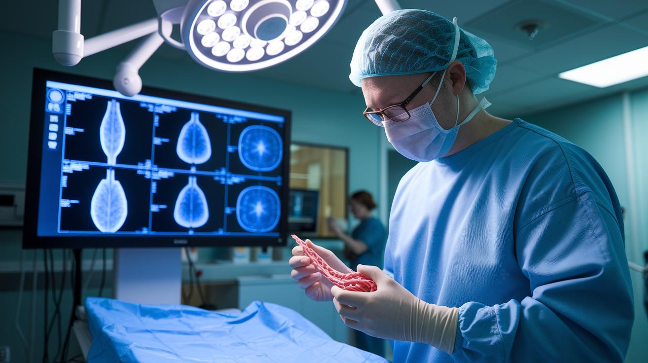
New 3D printing techniques are making surgery planning a lot more precise. For example, Materialise Mimics Innovation Suite takes CT scans (like detailed X-ray photos) and turns them into exact printed models of blood vessels, accurate to 50 microns. Imagine a surgeon looking at a real, three-dimensional map of a patient’s blood flow that can cut repair times for an aortic aneurysm by over two hours. This tech not only streamlines planning but also boosts confidence in personalized treatments.
Radiomics, a way to study disease through imaging, is also changing the game for tumors and biomarkers. The QIBA Profile v3.0 sets 127 standardized features (think of it as a detailed pattern guide) for various tumor types. And then there’s Visage 8.1 software, which can predict PD-L1 status in non-small cell lung cancer with an 82% match to traditional lab tests. This means doctors get a much clearer picture of a patient’s prognosis.
When you combine these imaging advances, you get a diagnostic tool that blends 3D printing with detailed data analysis. Now, doctors can see both a physical 3D structure and the numbers behind it, giving them a sharper view of a patient’s condition. This mix of visual and analytic information sets the stage for smart, tailored surgical plans and treatment choices.
Optimizing Radiology Workflows with Cloud and Sustainable Imaging Infrastructure
Cloud technology is really shaking up how radiology works by making it simpler to share data and by cutting costs. Take the RadioLance cloud teleradiology platform, for example, it runs nearly all the time (97% uptime) and can process CT scans in just 12 seconds. It even uses a secure method (blockchain-encrypted DICOM sharing) to move image files quickly and safely. Imagine being able to send scan data across different countries almost instantly, it’s a game changer for global diagnostics.
Another cool development is the rise of green technology in medical imaging. For instance, the GE SIGNA PET/MRI is designed with the planet in mind. It uses recycled magnets (made from rare-earth materials) and liquid nitrogen cooling, which means it avoids using helium. Despite these eco-friendly tweaks, it still delivers the strong 3 T field strength needed for top-notch images. These smarter, sustainable choices not only help the environment but also lower the running costs of these advanced systems.
There’s also a big push to streamline radiology with smart digital tools. Combining PACS and RIS systems (the setup for handling medical images and reports) has cut report turnaround times by half. Plus, interactive dashboards now give radiologists real-time updates on cases and radiologic data. This kind of integration makes workflows simpler, boosts productivity, and supports quicker, more informed decisions, paving the way for better patient care.
Future Perspectives on Medical Imaging Innovations
Medical imaging is on the brink of exciting changes that will reshape how radiology is practiced. New payment plans, smarter tools like augmented reality in surgeries, and a growing use of AI are gearing up to improve how doctors diagnose patients.
Value-Based Reimbursement Models
Payment methods are being revamped to reward precision. For example, under CMS 2025 MIPS, 35% of what radiologists earn now depends on clear imaging outcomes. This change pushes professionals to pay closer attention to the details in their images, which in turn fuels the creation of better imaging tools. Imagine a system where clearer pictures mean better funding for patient care.
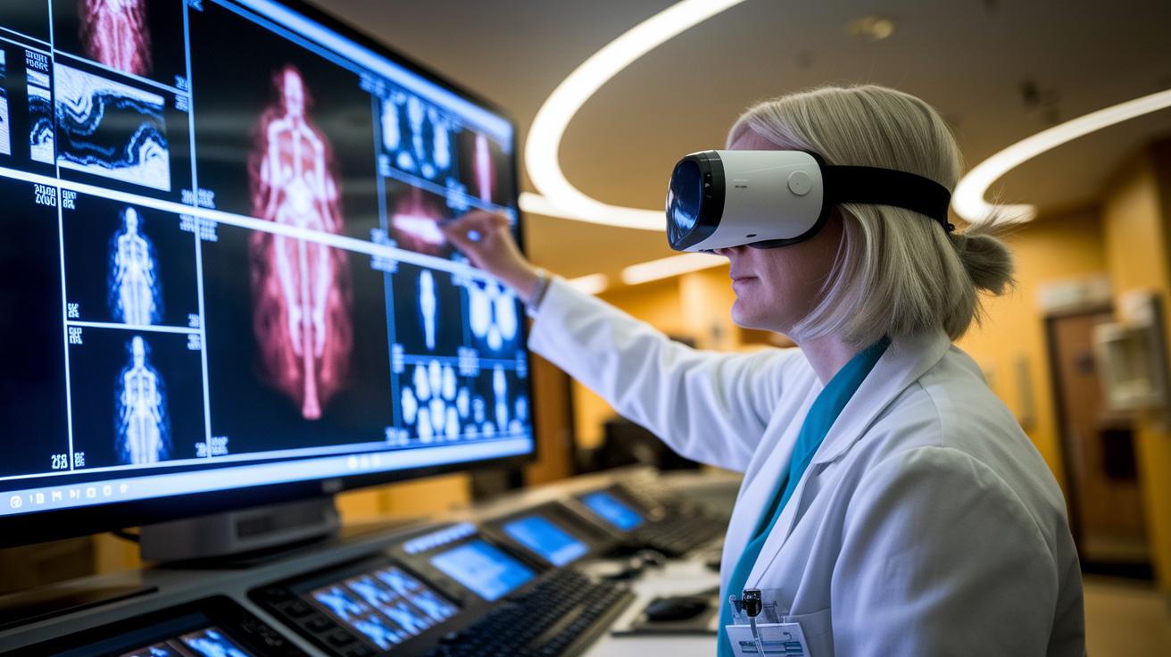
Augmented Reality in Interventional Imaging
Augmented reality is starting to change how surgeons work during operations. Early results show that AR-supported procedures can reduce operation times by 25%. Picture a surgeon seeing helpful imaging data right on the patient's body during a procedure, it makes the whole process smoother and more accurate. This tech is set to give doctors a real-time boost when they need it most.
Projected AI Adoption Rates
Experts predict that by 2030, AI will help with 70% of reading diagnostic images. Tools like advanced neural networks (smart computer programs that learn from data) will soon be everyday partners in reviewing scans, speeding up the diagnosis and making them more consistent. This expected shift is likely to accelerate the transformation of how medical imaging is done across the field.
Final Words
In the action, the article showcased how breakthrough technologies are transforming how we see diagnostics. It covered advanced scan systems, AI-powered imaging, and state-of-the-art 4D and hybrid methods that elevate patient care. The discussion also touched on how personalized diagnostics and smart cloud setups streamline workflows while driving down costs. Medical imaging innovations continue to spark rapid changes, offering promising tools that brighten the future of healthcare. The outlook remains positive as cutting-edge tech redefines our approach to diagnostics.
FAQ
Q: What does the artificial intelligence in medical imaging PDF include?
A: The artificial intelligence in medical imaging PDF outlines practical research, real-world applications, and technical details on using AI to enhance how doctors interpret images.
Q: What topics are covered in an AI in medical imaging research paper?
A: The AI in medical imaging research paper covers advancements in diagnostic tools, case studies of machine learning models, and comparative studies between traditional and AI-powered imaging systems.
Q: How does AI in medical imaging and diagnostics work?
A: AI in medical imaging and diagnostics works by using advanced algorithms, like convolutional neural networks, to analyze scans quickly and accurately, assisting clinicians in spotting disease signs early.
Q: What are some examples of AI in medical imaging?
A: AI in medical imaging examples include tools that match expert radiologists in detecting lesions, automated segmentation methods that reduce analysis time, and handheld ultrasound devices offering cost-effective screenings.
Q: Who are some AI medical imaging companies?
A: AI medical imaging companies include innovators that develop cloud-based diagnostic platforms and specialized software integrating deep learning to improve scan analysis and workflow in radiology.
Q: What are the benefits of AI in medical imaging?
A: The benefits of AI in medical imaging include improved diagnostic accuracy, faster image interpretation through automation, and early disease detection using predictive analytics, all of which support better patient outcomes.
Q: What does an AI in medical imaging course offer?
A: An AI in medical imaging course offers lessons on machine learning fundamentals, practical applications in scan analysis, and hands-on examples to help learners solve real imaging challenges in modern healthcare.
Q: How is salary determined in the field of medical imaging technology?
A: Medical imaging technology salary varies based on specialized skills in advanced diagnostic methods, experience with cutting-edge imaging tools, and expertise in optimizing radiology workflows.