

Have you ever imagined getting a new organ printed just for you? Thanks to 3D bioprinting, that idea is closer than you might think. This innovative process builds living tissue one layer at a time, kind of like putting together a colorful puzzle made of tiny cell pieces. With over 1,000 studies backing its promise, scientists are moving toward using printed cells for drug tests and tissue repairs in everyday care. In this article, we explore five exciting breakthroughs that could transform how patients are treated and even lead to custom therapies made just for your body.
Fundamentals of 3D Bioprinting in Medicine
3D bioprinting in medicine is an exciting process that uses special bioinks made from natural materials mixed with living cells. It works a lot like following a digital recipe, where the printer builds tissues layer by layer, kind of like putting together a living jigsaw puzzle, with each cell fitting perfectly to mirror real human tissue.
A really cool part of this technique is that scientists can print tissue models directly into the tiny wells used for experiments. This direct approach helps cut down on waste and makes everything more precise. Imagine printing a small patch of skin for a wound-healing test, each printed layer adds up to a piece of tissue that acts just like the real thing.
More than 1,000 studies show that 3D bioprinting speeds up drug testing in a way that’s kinder and more ethical compared to older methods. Beyond speeding up tests, this technology is also opening doors to create functional tissues and even organs that could be used in transplants. By combining smart materials with living cells, 3D bioprinting offers fresh opportunities for patient care and new research that pushes the boundaries of medicine.
Every layer built in this process is carefully made to form a living construct that could one day lead to personalized treatments. By merging digital blueprints with natural biology, this breakthrough technology is set to transform how we discover new drugs and develop regenerative therapies.
3D Bioprinting Workflow for Medical Fabrication
Pre-Bioprinting Stage
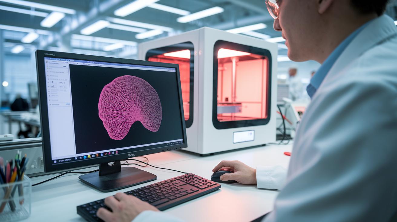
We start by creating a digital 3D model with a program like DNA Studio. This model acts like a map for the printer, showing exactly where each layer should go. Think about it: before making real living tissue, scientists first build a digital plan that mirrors nature’s amazing details. Engineers use different file types and design methods to capture every curve and tiny detail of the tissue design. This digital blueprint is key to making sure the following steps match the intended biological form.
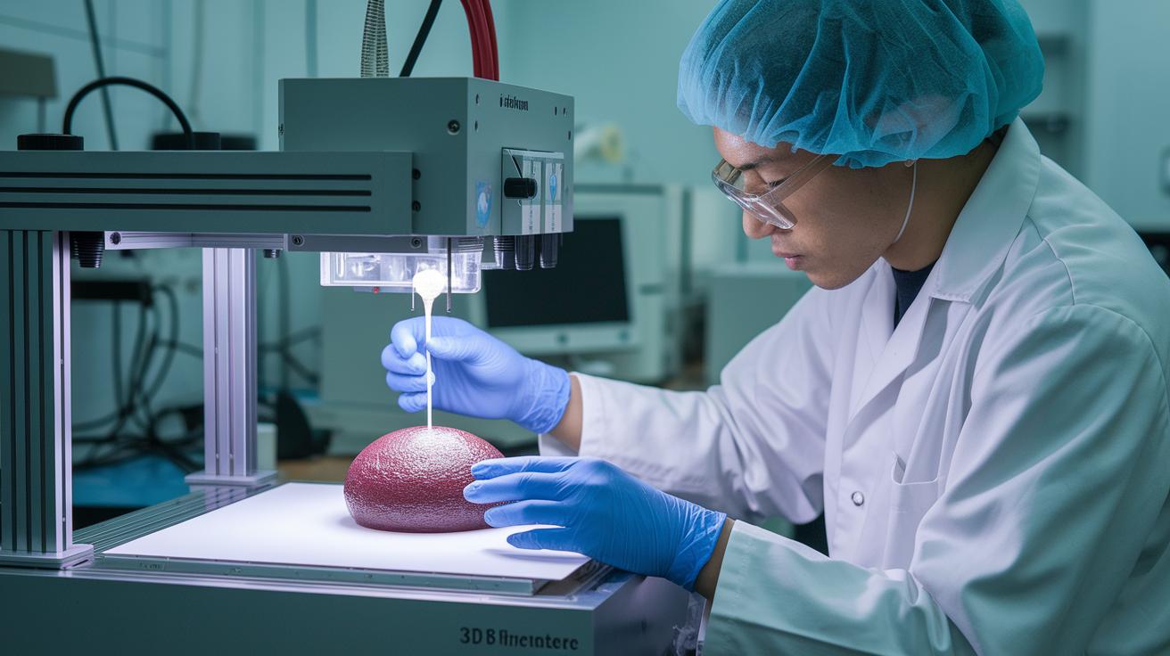
Bioprinting Stage
Next, the bioprinter steps in with impressive precision. This machine works at a 10 µm resolution (meaning it can place dots as small as 10 micrometers) and features special coaxial print heads with six changeable nozzles. The nozzle sizes vary from 18 to 34, each chosen to match the thickness and flow of the bioink (a mix that carries living cells). Picture it like piecing together a puzzle, with each drop of cell-filled material falling exactly into place. These careful settings help ensure that the tissue is built accurately and maintain high quality during every print.
Post-Bioprinting Stage
After printing, the tissue goes through a stabilization process using either ionic crosslinking or UV curing (using ultraviolet light to harden the structure). This helps the fabric hold its shape and work properly. Then, the printed structure is placed in an incubator, a controlled environment where conditions help the cells grow and the tissue mature. This final step is crucial for turning the fresh print into a functioning model that can be used for research and, possibly, future medical treatments.
Bioinks and Bioprinter Hardware Essentials
In medical 3D bioprinting, choosing the right bioink is super important. Scientists often use three main types: alginate, collagen, and GelMA. Alginate is a natural sugar (polysaccharide) that acts a bit like glue, helping to stabilize bone bandages and repair cartilage. Collagen, a key protein that supports our body’s structure, is used to make tissue models for skin grafts and to speed up wound healing. GelMA, short for gelatin methacryloyl, finds its role in methods using UV light to build tiny vascular channels. Did you know that just a tiny droplet of alginate can hold cells together much like glue?
Modern bioprinters come packed with features to handle a variety of bioinks. Their hardware is designed to fine-tune temperature and pressure, making sure each cell-laden mix does exactly what it needs to do. These machines can also work with different tissue-specific blends, allowing layer-by-layer building of incredibly detailed structures for personalized treatments.
The flexibility of bioinks and the compatibility of these machines are big moves toward more accurate tissue models. Here’s a quick reference guide to help you remember the basics:
| Bioink Type | What It’s Made Of | Medical Uses |
|---|---|---|
| Alginate | Natural algal sugar | Bone bandages, cartilage repair |
| Collagen | Main body protein | Skin grafts, wound healing |
| GelMA | Gelatin methacryloyl | Building tiny vascular channels |
Medical Applications of 3D Bioprinting: From Models to Transplants

3D bioprinting is changing the way we treat injuries and wounds in healthcare. Researchers are using printed patches that work like natural bandages to cover wounds. In one study, scientists printed layers of bioink filled with skin stem cells directly onto wounds in mice. Within just four weeks, the wounds grew new hair follicles and skin parts, much like a tiny patch of living bandage healing itself, even though there was just a small drop in the number of active cells.
Other cool projects are pushing things even further. For example, a team at Penn State used a special printing method that uses gentle suction to create a breast cancer tumor model with its own tiny blood vessels. This model not only looked a lot like real tumors but also reacted to treatments like chemotherapy the way true cancers do. It’s like having a mini lab inside the body to test new treatments and tailor them just for you.
At Brigham and Women’s Hospital, scientists are building sugar-based stents that help reconnect blood vessels during surgery. These smart stents dissolve on their own once blood starts flowing normally again, and they can be tailored to match each patient’s veins. This clever design helps cut down on long-term complications and shows promise in repairing organs in the future.
All of these breakthroughs are part of a bigger shift in medicine, moving from old-school treatments to living, tailor-made solutions. By using special bioinks and precise, high-tech printers, doctors might soon rely less on donor organs. It’s an exciting step toward more natural, efficient healing processes in clinics everywhere.
Clinical Impact and Challenges of 3D Bioprinting in Medicine
Over 100,000 patients in the U.S. wait for an organ transplant, and emergency rooms handle about 130 million visits each year. Early tests even printed bone and soft tissue onto a rat’s skull, proving the idea works on a small scale. Picture this: a piece of bone printed just right for a rat’s skull, showing that nature can be mimicked, even in tough conditions.
Even with these encouraging results, there are still obstacles to overcome. One big challenge is keeping the cells alive during printing, if too many cells drop off, the tissue might not work as it should in the body. And then there's the task of building tiny blood vessel networks (think of them as mini highways that nourish the tissue) that keep the printed tissue healthy.
Safety is a top priority when printing living cells and fitting implants into the body. Researchers are busy testing ways to make sure the process is safe without losing the tissue’s function. At the same time, creating standardized rules and overcoming regulatory puzzles, along with tackling ethical questions, adds to the complexity of the work.
Government bodies and research teams are working hand in hand to set clear guidelines. Next, cracking the code on vascular networks and implant integration will be key to bringing these exciting breakthroughs from the lab into everyday medical care.
Future Prospects for 3D Bioprinting in Medicine
The medical field is buzzing with excitement, thanks to over 1,000 peer-reviewed studies and all the teamwork among engineers, biologists, and doctors. Researchers are diving into ways to replace organs by perfecting printing methods that could eventually build entire organs layer by layer. Just imagine a printer working right at the spot where the tissue is needed, offering a faster, more personalized solution for patients.
Another exciting breakthrough is the automation of tissue fabrication. Scientists are crafting tiny, organ-on-chip systems that mirror how real tissues work, which helps with drug testing in real time. This setup not only gives us human-like data but could also reduce the need for animal testing. Digital twin tissue models are also on the rise, think of them as digital replicas of a patient’s tissue that doctors can use to plan surgeries and see how new treatments might play out.
New fabrication techniques are changing the way we build tissue constructs too. By mixing in nanotechnology (tiny machines at work), researchers hope to print tissues with built-in sensors that monitor their function continuously. Likewise, methods like bioreactor-assisted tissue growth are paving the way for steadier and more efficient cell development.
All these innovative ideas are sparking research that bridges the gap between groundbreaking lab work and real-world medical treatments. In the near future, combining digital blueprints, automated printing, and smart monitoring could bring experimental models much closer to patient-ready therapies, marking a promising new era in personalized healthcare.
Final Words
In the action, we explored how 3D bioprinting in medicine works, from crafting digital models to printing living tissues and overcoming safety challenges. The blog post broke down every step of the process, discussed bioinks and printing hardware, and looked at real-world applications like tissue repair and transplant research.
We also highlighted emerging trends that forecast exciting advancements in regenerative care. The progress in 3D bioprinting in medicine inspires an optimistic future for patient care and innovation.
FAQ
What is 3D bioprinting and what is bioprinting?
The 3D bioprinting is a method that uses bioinks (a mix of biomaterials and cells) to build living tissue layer-by-layer from digital designs, creating models for research and clinical applications.
What is 3D bioprinting used for in healthcare and medicine?
The 3D bioprinting is used to create tissue models, skin grafts, bone bandages, and other constructs that aid in drug testing, wound repair, and eventually, transplant procedures.
What are examples of 3D bioprinting in medicine, including bioprinting of organs?
The 3D bioprinting examples include printed skin grafts, bone bandages, vascularized tissue models, and emerging techniques aimed at producing transplantable organs and functional tissue constructs.
How does bioprinting work?
The bioprinting works by depositing bioinks in successive layers according to a digital blueprint, involving design preparation, high-precision printing, and post-processing methods like crosslinking to stabilize the tissue.
What does 3D bioprinting in medicine online refer to?
The 3D bioprinting in medicine online refers to digital platforms and resources that showcase research, demonstrations, and real-world applications of 3D bioprinting technologies in healthcare.
What is the FDA approved 3D printing drug?
The FDA-approved 3D printing drug is a medication manufactured using additive processes with regulated standards to guarantee safety and efficacy, as documented by official FDA releases.
How long until we have fully functional 3D-printed organs?
The timeline for fully functional 3D-printed organs is still developing, with ongoing research suggesting that it may take several more years before these constructs are ready for routine clinical use.